Ureteral obstruction, especially in malignant
diseases
Updated:
July 2017
Anatomy
The urinary tract including
the ureters allows urine flow from the kidneys to the bladder. Ureters
are active members. Their wall contains muscle fibers which contract. A
wave of contraction is triggered in the kidney calyx and spreads like a
wave throughout the pelvis and all of the ureter.
The ureter is very thin.
If there is a severe suffering on a portion of the ureter, it shrinks circumferentially
and obstructs its own light. This wall destroyed will never heal. It must
be removed (cut and paste) or occupied for life with a "plastic" tube.
Diseases of the
ureter
The pathology of the ureter
is large and complicated. Diseases of the ureter are very interesting.
I suggest you to explain by the following short video "Autopsy of a obstructive
double-pigtail tumorstent" and by the page below, why it's a challenge.
Autopsy of a obstructive
double-pigtail tumorstent
When the ureter is obstructive
and it is not a stone or a ureteropelvic junction, it is essential to discover
the mechanism of the obstruction.
The mechanism is sometimes
suspected by the history of the patient (malignant diseases, surgical treatment
for a tumor, radiotherapy). But sometimes it origin is only discovered
during the drainage of the kidney.
The
origin of the obstruction
The ureter may be
the cause of his own obstruction. If a tumor growth in the wall, it clogs
progressively its light. The tumor of the ureter may be malignant or not.
A dilation of the kidney appears often without any pain. The tumor can
bleed and thus be discovered by red urine (hematuria). The tumor can suddenly
be obstructive by abundant bleeding and causes severe pain with renal colic.
The ureter may be
crashed by a foreign process. Processes can be of different origins:
- They can be benign
(not malignant). This is the case of endometriosis, or retroperitoneal
fibrosis, or traumatic by difficult surgery around the ureter.
- They can be malignant.
This is the case of cancers that directly reach the ureter (colon, uterus),
or via a node cancer (metastasis prostate, bladder, ureter, colon, uterus),
or malignant retroperitoneal fibrosis of the breast. In fact, breast cancer
metastasis can reach this region and can realize an extremely rigid matrix
crushing the ureters.
Obstruction
of the right ureter by prostate cancer nodes.
Side effects of the obstruction
If the ureter is gradually
dilated, no pain is observed. But a suddenly edema of the tumor may obstruct
the kidney and causes severe pain with renal colic.
The silent obstructions may
have important functional side effect: complete destruction of one or both
kidneys.
The suddenly obstruction
of one or both ureters can have serious or critical side effect: pain,
fever, sepsis, renal failure, death.
Treatments
The treatment is to restore
urinary flow and prevent renal destruction.
In case of ureteral
tumor, tumor must be removed with all ureter below the tumor. There are
often tumor grafts on the entire length of the ureter. Thus, kidney is
loss because no ureter exists to connect to the bladder.
In case of ureteral
tumor compression or ureteral stricture after radiotherapy, a double-pigtail
stent is currently inserted to bypassed the obstacle.
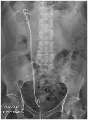
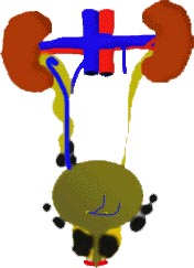
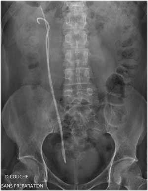
Drainage of the right
ureter by a strong (reinforced) double-pigtail stent.
But insertion of double-pigtail
stent in a compressed ureter is not equivalent to success.
This concept is
CRUCIAL.
The stent will be obstructive
quickly ...
The tumor is sometimes very
compressive and breast tumors are the hardest tumors. Then, reinforced
double-pigtail stent are marketed with reinforcing tube, or metal coils.
With the tumor, the stent
must be very strong against compressive and kinking forces. It needs allowing
patency. There may be deposits by stagnation and particle-forming by infection.
These particles are embedded in the wall and decrease the light of the
stent. The compression of the stent reduces the obstructive light of the
stent and, full obstruction of the stent may occur.
The suddendly obstruction
of the stent may have serious or critical consequences: pain, fever, sepsis,
renal failure, death. The silent obstructions of the stent may have functional
consequences by destroying the kidneys. This obstruction occurs in a tired
patient and may kill this patient. This death is not directly induced by
chemotherapy or cancer but just by the effects of the stent's obstruction.
Finally, repeated stent's
exchange and poor tolerance of the stent affect patients' quality of life.
For this reason, it seems
to me essential to place the most "efficient" stent and detect any incompetence
of the stent. This strategy require that the urologist frequently exchanges
the stent, even if it is designed for one year.
These tumorstents "special
for tumor" whatever their composition are not be efficient more than 6
months. 40% are obstructive within 3 months and sometimes in less than
a week. In the latter case, it is crushed. Everyone agrees that we must
find an optimal type of stent with adequate permeability and resistance
[références
1-3]
In case of emergency or impossibility
to insert a double-pigtail stent, a nephrostomy (through the back) is possible.
But this drainage is uncomfortable and unstable. The probe may move out
of the kidney.
Right nephrostomy tube
draining urine by the back.
Personally, I struggle to
find an original solution. I have to understand why the stent does not
work (twisted ? ), or be obstructed too quickly (crashed ?). Then, I have
to adjust the drainage means.
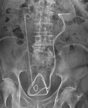
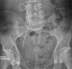
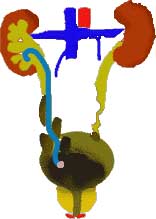
For example, I can
use 2 large stents of 9 French side by side to stiffen and verticalize
ureter.
Insertion of large stents
to drain a tortuous left ureter.
For example, I can
use 1 large stent 8 French with a reinforced double-pigtail stent.
Insertion of large stents
for draining heavily crushed ureters.
The patient must return to
the operating room but finally urine patency is effective and patient's
life is better.
Of course, the amount of
material in the bladder may induce bladder irritation. In some cases, these
stents can be cut and become reinforced JFil®
stent and improve the patient's life comfort [références
4-7].
Reinforced
JFil® stent using the stent into the tumor and a single thread
in the rest of the ureter and the bladder.
Reinforced
JFil® stent using the stent in the tumor. The thread in the
bladder is not visible on the X-Ray.
In other cases, the lower
part of the ureter is obstructive and the use of a stent with a thread
is not possible. Then, I cut the stent to eliminate purely bladder part
and thus reduce bladder irritation. An end piece is put at the bottom of
the stent. These procedures have been made more than 40 times and the results
are very encouraging [référence
8]. This new stent improves patient
comfort of life and I am looking for industrial partner.
A clinical trial is now readfy to start by Dr. B. Vogt.
Development
of an effective tumorstent should be of concern
Reinforced
JClip stent using the stent into the tumor and an end piece in the
bladder.
Reinforced
JClip stent using the stent in the tumor. The end piece in the bladder
is not visible on the X-Ray.
Surveillance of
patient
In case of kidney pain, unexplained
fever or renale failure, we must realize sonography of the kidneys and
compare pelvic dilation.
My advice is to check sonography
each 2-3 months.
Medical
studies
1. Yossepowitch O et al.
Predicting the success of retrograde stenting for managing ureteral obstruction.
J Urol 2001, 166: 1746-9.
2. Chung SY et al. 15-year
experience with the management of extrinsic ureteral obstruction with indwelling
ureteral stents. J Urol 2004, 172: 592-5.
3. Hendlin K. In vitro evaluation
of ureteral stent compression. Urology 2006, 67: 679-82.
4. Vogt B et al. Sondes JFil
et MiniJFil : progrès décisifs dans la tolérance des
sondes urétérales et propriétés inattendues
du fil urétéral. Prog Urol 2014, 24: 441-50. doi: 10.1016/j.purol.2013.12.007.
http://www.sciencedirect.com/science/article/pii/S1166708714000189
5. Vogt B et al. Changing
the double pigtail stent by a new suture stent to improve patient quality
of life. A prospective study. World J of Urology 2015, 33: 1061-8. doi:
10.1007/s00345-014-1394-2. (Open Access)
link.springer.com/article/10.1007%2Fs00345-014-1394-2
6. Vogt B et al. Improving
the quality of life of patients with ureteral malignant obstruction. J
Palliat Care Med 2014, 4: 196. doi: 10.4172/2165-7386.1000196. (Open Access)
omicsgroup.org/journals/ArchiveJPCM/articleinpress-palliative-care-medicine-open-access.php
7. Vogt B et al. Changing
the shape of the double-pigtail stent to attenuate urinary stent's symptoms.
J Palliat Care Med 2014, 4: I101. doi: 10.4172/2165-7386.1000I101. (Open
Access)
omicsgroup.org/journals/ArchiveJPCM/articleinpress-palliative-care-medicine-open-access.php
8.
B. Vogt et al. Improving Comfort of Patients with Ureteral Obstruction
and Malignant Disease Should Be of Concern. J Palliat Med. 2016 Nov;19(11):1132-1133.
OpenAccess. DOI: 10.1089/jpm.2016.0276